.gif)

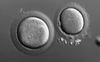
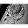
OocytesIVF Image Database - Oocytes31. 'Giant' oocyte
Views: 6830
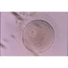
32. GV-stage oocyte
Views: 9892
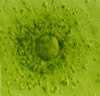
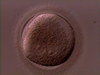
33. Oocyte
Views: 5304
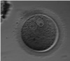
34. Two GVs in One Prophase I Egg
Views: 5105
35. Abnormal Zona Development
Views: 4876
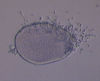
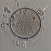
36. Germinal Vesicle
Views: 5325
37. Human Empty zona-cumulus-corona cell complex
Views: 8126
38. Zona-Free Oval Shaped Oocyte
Views: 5437
39. Immature oocyte with two germinal vesicles
Views: 7044
40. An egg with cellular fragment in Zona Pellucida
Views: 7319
|