

IVF NewsNews: Discovered: The starting pistil for sperm
Rose Palmer 09 February 2010
Scientists from the University of California in San Francisco have identified the mechanism by which sperm start swimming towards the egg when they enter the female reproductive system. The discovery could lead to drugs that boost male fertility and new forms of contraceptives. The finding was reported in Cell. It has been known for a while that a sperm's level of activity is affected by a change in internal pH, but the exact mechanism that regulates swimming was unknown. To investigate, Dr Yuriy Kirichok and his team used a technique called patch clamping to record proton movement across the cell membrane of sperm. They discovered that there was an abundance of Hv1 proton channels in the tails of the sperm. These act as a pore in the outer membrane of the sperm cell, extrude protons and are responsive to changes in the levels of zinc and pH outside of the cell. The uterus has a pH concentration one thousand times higher than semen, and this triggers the Hv1 channels to open. Extrusion of protons makes the environment within the sperm more alkaline and this, in turn, causes the sperm to start swimming. High concentrations of zinc, as found in semen, inhibit the Hv1 channels, preventing them from opening too soon. The levels of zinc are lower in the fallopian tubes and this may trigger an extra spurt of swimming power as the sperm nears the egg. Dr Kirichok said: 'What we're very excited about is that we've found the molecule that elevates sperm intracellular pH and we've found how that molecule is activated'. The researchers found that a compound called anandamide, which is found in high levels near the egg, also causes the channels to open. The compound is similar to the active ingredient in cannabis and it is possible the drug may mimic the effect. This could explain the link between cannabis use and poor fertility in males. The finding could lead to new forms of contraception. Dr Kirichok said the channel could be exploited by a drug which hampers proton release, leaving the sperm unable to swim. He said: 'All of these events are essential to fertilisation - you can imagine now that we know the molecule responsible we could block it to prevent activation and fertilisation as a kind of male contraception'. It may now also be possible to find a way to improve the sperm mobility of men who have fertility problems. [ Full Article ]  
News: Australia considers lifting ban on sex selection
Nishat Hyder 16 March 2010
The Australian federal five-year moratorium on the use of gender selection technology in IVF (in vitro fertilisation) treatment for so-called 'social' reasons ends this year, reopening this controversial debate. The Australian health watchdog, the National Health and Medical Research Council (NHMRC), confirmed that that it will be conducting a review of this issue, beginning within the next few months, after the completion of its ongoing review of the Research Involving Human Embryos Act and the Prohibition of Human Cloning for Reproduction Act. At present the use of gender selection technology - specifically, selection by using PGD (preimplantation genetic diagnosis) - is only allowed where parents carry a serious genetic disorder that can be passed on and detected through gender. The social use of gender selection was banned in 2004 by federal legislation, and earlier in 1998 by state legislation in Victoria. How the NHMRC will choose to act post-review is the subject of much speculation. It is important to note that any legislation introduced by this body is federally binding and would override state legislation. An alternative route would be for the NHMRC to introduce guidelines allowing gender selection on non-medical grounds, therefore leaving the legislative decisions about what specific grounds might be included within this - or not - to individual state and territorial governments. Such course of action would create a situation where the choice of gender selection is legally available in some Australian states, but not in others, and might lead to 'inter-state fertility tourism'. Fertility tourism for gender selection purposes is by no means a new phenomenon. The Australian newspaper, the Herald Sun, revealed in December 2008 that many Australian couples were travelling to the US or Thailand for fertility treatment where they are free to choose the gender of their baby. The use of gender selection technology for non-medical purposes such as 'family balancing' or because of cultural influences is controversial on ethical and religious grounds. However, there has been a strong lobby from within the medical profession in favour of allowing this technology for social reasons. Professor Gab Kovacs, an IVF pioneer, is leading the lobby: 'If I am prepared to pay for it out of my own pocket so it is not the community paying, I can't understand why that should be forbidden,' he said, adding: 'It might even be in the interests of the child. If a couple so badly want a boy or a girl that they are prepared to go through IVF and gender selection then maybe, if they had the child naturally and it was the wrong gender it may not be looked after as well'. Professor Kovacs further points out that at $10,000 - $15,000 only extremely determined couples will actually use gender selection technology. Dr Michael Chapman, a senior infertility specialist at IVF Australia, echoed this sentiment: 'Very few patients I see ask to choose the sex of their child - less than five per cent'. Across Australia many doctors are voicing their support for freedom of choice when it comes to gender selection. Some however, such as Dr Lyndon Hale, chairman of Melbourne IVF, are tempering their support. Although Dr Hale is in favour of giving parents the choice to use gender selection technology, he believes that this should only be allowed where there is a significant benefit for both the parents and the child in doing so. [ Full Article ]
Article: Improved development of mouse and human embryos using a tilting embryo culture system
RBM Online 28 March 2010
Koji Matsuura a,b, Nobuyoshi Hayashi c, Yuka Kuroda a,b, Chisato Takiue c, Rei Hirata c, Mami Takenami a, Yoko Aoi c, Nanako Yoshioka c, Toshihiro Habara c, Tetsunori Mukaida d, Keiji Naruse a,* a Cardiovascular Physiology, Graduate School of Medicine, Dentistry and Pharmaceutical Sciences, Okayama University, Okayama, Japan; b Research Core for Int erdisciplinary Sciences, Okayama University, Okayama, Japan; c Okayama Couples Clinic, Okayama, Japan; d Hiroshima HART Clinic, Hiroshima, Japan * Corresponding author. E-mail address: [email protected] (K Naruse).
Dr Keiji Naruse graduated from Nagoya University School of Medicine in 1988 (MD) and received his PhD in medicine from Nagoya University in 1992. He was an associate professor at Nagoya University from 1999 to 2005 and is currently a chairman and professor of Cardiovascular Physiology at Okayama University Graduate School of Medicine. He was a visiting professor at Harvard Medical School from 1998 to 2001. He has been working in the fields of mechanobiology of circulation, reproduction, and sensory systems. Abstract Mammalian embryos experience not only hormonal but also mechanical stimuli, such as shear stress, compression and friction force in the Fallopian tube before nidation. In order to apply mechanical stimuli to embryos in a conventional IVF culture system, the tilting embryo culture system (TECS) was developed. The observed embryo images from the TECS suggest that the velocities and shear stresses of TECS embryos are similar to those experienced in the oviduct. Use of TECS enhanced the development rate to the blas- tocyst stage and significantly increased the cell number of mouse blastocysts (P < 0.05). Although not statistically significant, human thawed embryos showed slight improvement in development to the blastocyst stage following culture in TECS compared with static controls. Rates of blastocyst formation following culture in TECS were significantly improved in low-quality embryos and those embryos cultured under suboptimal conditions (P < 0.05). The TECS is proposed as a promising approach to improve embryo development and blastocyst formation by exposing embryos to mechanical stimuli similar to those in the Fallopian tube. RBMOnline © 2009, Reproductive Healthcare Ltd. Published by Elsevier Ltd. All rights reserved. Declaration: Keiji Narusee launched the bio-venture company, Strex Inc., in 2003 and serves as a Chief Scientific Officer. The other authors report no financial or commercial conflicts of interest. KEYWORDS: blastocyst, embryo development, mechanical stimuli, shear stress, tilting embryo culture system Introduction Mammalian embryos are transported to the uterine cavity through the Fallopian tube during cell cleavage, blastomere and blastocyst development (Eddy and Pauerstein, 1980; Halbert et al., 1976). In conjunction with ciliated epithelium, the Fallopian tube acts as a peristaltic pump due to phasic contraction of the smooth muscle in the wall to transport the embryo (Lyons et al., 2006; Zervomanolakis et al., 2007). Based on the movement of cilia and the similar size between the tubal lumen of ampulla and isthmus (Table 1) and the diameter of the embryo (0.1 mm), fertilized oocytes may be subject to a mechanical influence from the Fallopian tube lumen, such as compression and shear stress from the tubal fluid. It is postulated that these mechanical actions in the Fallopian tube might play an important role in embryo development. However, conventional in-vitro static culture conditions do not mimic mechanical stimuli to embryos. Providing mechanical stimuli to developing embryos in-vitro, similar to that experienced in the Fallopian tube, may improve development. 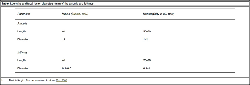 Table 1. Click on image for full size. From the 1980s, some groups reported in-vitro non-static culture results (Nagai et al., 1993; Staigmiller and Moor, 1984). However, these reports did not discuss the precise effect on embryo development from cleaved embryo to blastocyst. Recent papers have reported that a microfluidic dynamic embryo culture system with media flow improved mouse embryo development (Cabrera et al., 2006). The report proposed the importance of the physical/mechanical environment on embryo development. Because the systems are complex to handle, clinical application of the culture system is quite difficult. In addition to ease-of-use, another consideration regarding mechanical stimulation during embryo development is avoiding excess stress. Excess mechanical stimuli would damage embryos. It was found that shear stress over 1.2 dyn/cm2 caused lethality within 12 h for blastocysts (Xie et al., 2006). Over-handling of embryos, such as excess pipetting, caused elevation of phosphorylated stress- activated protein kinase and may cause rapid transient changes in hundreds of proteins and mRNA (Xie et al., 2007). In order to construct a dynamic culture system that can apply a mechanical stimulus and can be easily adapted to conventional static culture platforms, the tilting embryo culture system (TECS) was developed. Placing a conventional culture dish or plate on a tilted plate makes embryos in culture move along the bottom of the dish by gravity. Animal model experiments using TECS were first conducted, for the in-vitro culture of mouse 2-cell-stage embryos. A subsequent study using thawed human embryos cultured on TECS was also performed. Materials and methods Observation of embryo motion and shear stress calculation To estimate the shear stress applied to embryos, mouse embryo motion in the microdrop on the tilting plate of a prototype TECS was observed by objective lens (20x) attached to the bottom of the tilting TECS plate. To estimate the flow velocity of media in the microdrop, this study observed the motion of the microspheres (0.5–5 μm diameter) in the microdrop covered with mineral oil by tilting inverted microscope (Eclipse, Nicon, Tokyo, Japan), particularly focusing on the motion of microspheres at the centre of the bottom where embryos in the microdrop were moving. When the microscope was tilted, the particles moved in the same direction as the leaning microscope. The average particle velocities, which were considered as the velocity of the medium in the microdrop, were calculated. Under the holding the tilt and static conditions, Brownian motion of the particles was dominant. Images of moving embryos and microspheres were recorded by a charge-coupled device camera connected to a personal computer. The frame rate of the recording was 30 frames/s. The observed maximum velocity of the embryos and the velocity of the medium were VE and VM, respectively. The velocity to estimate maximum shear stress (VSS) was calculated from the difference between VE and VM. VSS =VE -VM The maximum shear stress applied to embryos during the tilting was calculated from the following equation: Shear stress = 6µrVSS =4µr2 = 6VSS dyn/cm2 where µ is the viscosity of the medium and r is the radius of the embryo. In an attempt to provide as accurate an estimate as possible regarding sheer stress experienced by embryos, this study attempted to determine the flow velocity of fluids using microspheres. The above equations are applicable in the flow condition. Generally, the effect of Brownian motion during flow can be neglected for the shear stress calculation. However, it would be difficult to determine accurate fluid velocity by particle image velocimetry. Although forces such as friction come into play with this approach, it provides a rough estimation for the final calculations. Embryo motion in extirpated mouse oviducts was observed as follows. Female ICR mice (8–12weeks old; Charles River Japan, Yokohama, Japan and Shimizu Laboratory Supplies, Kyoto, Japan) were injected with 5 IU pregnant mare’s serum gonadotrophin (Aska Pharmaceutical, Tokyo, Japan), followed by 5 IU human chorionic gonadotrophin (Aska Pharmaceutical) 48h later and mated with males. Successful mating was determined the following morning by the detection of a vaginal plug. Two days after the mating, the oviduct was extirpated with embryos and the oviduct was sandwiched between a cover slip and a glass slide to observe the embryo in the oviduct. The velocity of the embryo in extirpated mouse oviducts was calculated from the movie of the embryo motion. Animal use protocols were approved by Okayama University animal investigation committee. Specification and motion programmes of TECS TECS (Strex, Osaka, Japan) is an electrical device with a power cord that is designed to be used in a humidified incubator. This device consists of a control unit (Figure 1A, upper) and a motor unit with a tilting plate (Figure 1A, lower). Four-well chambers and/or dishes are set on the plate. The TECS motor unit is controlled and DC-powered by a cord connected to the control unit through an access hole in the incubator. If the incubator does not have an access hole, a flat-type cable extends tightly through the door of the incubator in order not to alter temperature, gas concentration and humidity inside the incubator. The motor unit is waterproof. The controller outside the incubator puts in the motion parameters.  Figure 1. Click on image for full size. A representative motion programme of TECS is shown in Figure 1B and C. The TECS can control the parameters of the uniform radial velocity (Vr), the maximum tilt angle (θr) and the holding time at the maximum tilt angle (Th). The tilting time (Tr) in seconds is calculated from 2θr/Vr. First, the plate is tilted to the positive maximum tilt angle (+θr) (M1). Second, the tilting plate is held for Th with no motion (M2). Third, the plate is tilted to the negative maximum tilt angle (−θr) (M3). Last, the tilting plate is held for Th with no motion (M4). The TECS motion cycle is summarized in Figure 1C. The cycle continues until the power is turned off. To apply conventional culture dishes/approaches to the TECS, spill out of the mineral oil should be prevented. The angles at which mineral oil begins to spill out are different according to the size of the dishes. The mouse embryo experiments used dishes of 35 mm diameter and maximum tilt of 20 degrees. However, in the human embryo study, the plate could not be tilted over 10 degrees due to the use of dishes of 60 mm diameter. The maximum tilt angle that caused mineral oil to spill out was surveyed and the limit of the maximum tilt angle was approximately 20 degrees in the microdrop setup. Furthermore, excess uniform radial velocity also induces spill out of the mineral oil. The minimum radial velocity at which the mineral oil spilt from the 35 mm dish was 240 degree/s when the maximum tilt angle was 20 degrees. The oil did not spill out when the tilt angle was 10 degrees. It is necessary to increase the maximum tilt angle and radial velocity in order to move embryos in the microdrop. However, this study set the maximum tilt angle and radial velocity so that they did not result in the spilling out of the mineral oil but still allowed the observation of embryo motion in the video rate recording. Thus, the maximum tilt angle was 10–20 degrees and the radial velocity was approximately 1 degree/s. Tilting mouse embryo culture Frozen 2-cell-stage embryos of ICR mouse (Arc Resources, Kumamoto, Japan) were thawed and cultured in 50 and 500 μl, respectively, of potassium simplex optimized embryo culture medium (Daiya Shiyaku, Tokyo, Japan). The culture medium was covered with mineral oil and incubated for 3 days in a humidified environment of 5% CO2 in air at 37°C. In the TECS group, the mouse embryos were cultured at a maximum tilt angle of 20 degrees with a holding time of 1 min. The plate was rotated at 1 degree/s to reach a total tilt of 20 degrees. To count cells in the mouse blastocyst, the cells were stained with Hochest 33342, as previously reported (Hardy et al., 1989; Mottla et al., 1995). The stained blastocysts were observed by confocal microscopy (FV-1000; Olympus, Tokyo, Japan) and a 3D image was constructed. Human embryo study The human embryo study used 3- to 11-cell-stage embryos frozen by the slow method 3 days after collection of the oocytes from May 2000 to December 2004 (Cohen et al., 1985) and extended the prospective study of 220 fertilized human embryos that were to be discarded with consent after pregnancy of patients. This study was approved by the ethics committee of the Okayama University Graduate School of Medicine. The frozen embryos were thawed with THAW-KIT 1 (Vitrolife, Gothenburg, Sweden) and the viability of the thawed embryos was approximately 80%. Once thawed, human embryos were cultured in a 20 ll microdrop of Global medium (LifeGlobal, Ontario, Canada) covered with mineral oil for 48 h. The thawed embryos with morphologically regular cleavage were divided so that there would be the same percentage of embryos with regular cleavage (33%) in both the TECS group and the control group. The viable cell number of the thawed human embryos was from three to 11. The dishes were placed on the tilting plate of TECS in a 50 l multi-gas incubator (ASTEC, Fukuoka, Japan). The thawed human embryos were cultured at a 10 degree tilt for 10 min. The radial velocity of the tilt was 1 degree/s (Vr). The blastocysts were morphologically evaluated according to the classification of Gardner et al. (2000). The cells in the human blastocyst were stained with Hochest 33342, the blastocysts were observed by confocal microscopy (FV-1000 Olympus, Tokyo, Japan) and a 3D image was constructed. Statistical analysis Chi-squared test and Student’s t-test were used to determine differences in the blastocyst development rate and in the number of cells in the blastocysts between the groups, respectively. A P-value < 0.05 was considered significant. ResultsObservation of embryos in motion by TECS and estimation of shear stress To obtain detailed information on the embryo motion, the blastomeres were observed on the TECS during tilting and holding. Figure 2 shows cropped images of the mouse embryos in motion. Between each 10 degree tilt (M1 and M3 in Figure 1B), the TECS plate was tilted at 1 degree/s. The motion of the medium was observed in M1 and M3 (Figure 1B). The embryo moved at a velocity of 1.5 mm/min on average (Figure 2A and B), which gave rise to shear stress of 7.0 x 10⁻³ dyn/cm2 at the bottom of the centre in the microdrop at a velocity of 0.1 mm/min. When the TECS plate was held at a 10 degree tilt (M2 and M4 in Figure 1B), the embryo slid at a velocity of 0.03 mm/min (Figure 2C, D), which gave rise to a shear stress of 1.5 · 10–4 dyn/cm2 in the medium, where the motion of medium at the centre of the bottom was neglected in the motion phases. These values are summarized in Table 2. 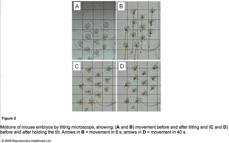 Figure 2. Click on image for full size. 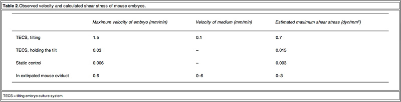 Table 2. Click on image for full size. Mouse embryo development Table 3 shows the blastocyst development rates from 2- cell-stage mouse embryos cultured under several conditions. As shown in Figure 3, the blastocyst development rate was 79% for 10 embryos in a 50 ll microdrop using the TECS, which was not significantly different to the static culture controls (75%). Because mouse blastocyst development rate is reported to be influenced by the number of embryos in the culture medium (Melin et al., 2009) and in order to enhance the statistical difference between TECS and control, the number of embryos in the microdrop were deduced and the medium volume-to-embryo ratio, which might simulate poor development condition and lead to low-quality human embryos, was increased. The blastocyst development rate for four–six embryos in a 50 ll microdrop was less than that of 10 embryos, although in this poor condition, TECS significantly improved the blastocyst development rate (TECS 59% (n = 145) versus control 46% (n = 151); P < 0.05). When 10 embryos were cultured in 500 ll medium, the blastocyst development rate decreased to 27% (n = 101) in the static control, whereas for those cultured in the TECS it was significantly higher (42% (n = 99); P < 0.05)). The number of cells in the blastocysts cultured using the TECS was (mean ± SEM) 77 ± 4 cells (n = 34), while that of the control was 66 ± 4 cells (n = 26), as shown in Figure 4. There was a significant difference in the average cell number between the two groups (P < 0.05). 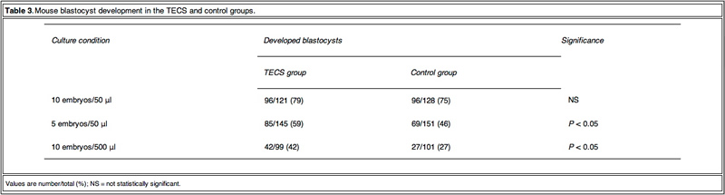 Table 3. Click on image for full size. 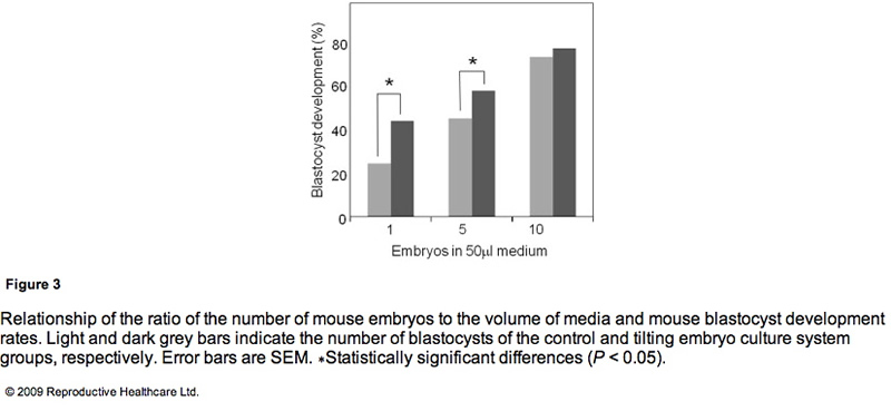 Figure 3. Click on image for full size.  Figure 4. Click on image for full size. Human embryo development As TECS enhanced blastocyst development rate in mouse embryos, experiments were then conducted on thawed human embryos. The development rates to the blastocyst stage of the TECS and control groups were 53% and 45%, respectively (Table 4). 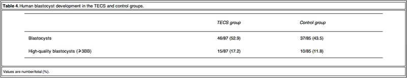 Table 4. Click on image for full size. Due to the insufficient sample number of thawed human embryos, the study investigated the cell number of the developed blastocysts instead of the blastocyst development rate. Figure 5 shows the comparison of average cell numbers in human blastocysts developed by the TECS and control groups. The mean cell number of developed blastocysts at day 5 by the TECS was 43±3 cells (n=24), while that of the control was 34 ± 3 cells (n = 18). There was a significant difference in the average cell number between the two groups (P < 0.05). 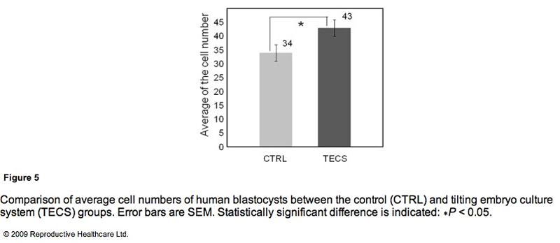 Figure 5. Click on image for full size. Discussion The Fallopian tube is a multifunctional organ, involved in receiving ovulated oocytes, providing a suitable environment for fertilization and early development and transporting embryos to the uterus. Here, light is shed on the mechanical properties of the Fallopian tube that might influence early development of embryos during culture. Early studies showed that the Fallopian tube is a mechanically active organ and may have influence on development due to: (i) shear stress by a tubal fluid flow; (ii) compression by peristaltic tubal wall movement; (iii) buoyancy; and (iv) kinetic friction force between embryo and cilia. Punctuated velocities of maxima from 0.39 to 1.8 mm/min have been observed in rat oviducts for microspheres emulating the size of embryos (Xie et al., 2006). Inappropriate culture conditions could be detrimental. Indeed, embryos sense shear stress and development is compromised (Xie et al., 2006, Xie et al., 2007). Because shear stress is a function of the velocity of the embryo and the flow and because the contribution is important, it is propose that the velocity of embryos should be made similar to those in the oviduct. Similarity in the velocities of the mouse embryos was found but those of other species were not observed. The velocities in-vivo and ex-vivo should be investigated for optimization. The observed velocity of mouse embryos on a TECS culture plate was of a similar order to those proposed in the oviduct. The embryo motion in the microdrops on the TECS plate relates directly to the plate motion. The observed embryo images on the TECS plate suggest that the velocities and shear stresses of embryos (Table 2) in the TECS plate and the oviduct are similar. This comparative experiment suggests that the TECS can apply physiological mechanical stimuli to mouse embryos. In the case of human embryos, the embryo velocity in the microdrop on the TECS plate was the same order (approximately 1 mm/min) as those of mouse embryos and below 1.2 dyn/cm2. Therefore, the TECS could apply physiological stimuli without an excess amount of shear stress that might cause damage to the embryos. Furthermore, the detrimental or beneficial shear stress would be different at each developmental stage and future experiments will address this. Embryo motion in fluid is affected not only by species differences but also the environment of the embryos. For example, when cumulus cells are coated with zona, embryos with the cells stick on the bottom of the dish. To optimize the parameters (θr and Vr) for each embryo condition, the relationship between the motions in fluid and physical characteristics of embryos (such as density, dimension and zona surface structure) should be considered. The TECS significantly improved development in low-quality human embryos and suboptimal culture conditions in mouse embryos. The significance of the improvement by the TECS was dependent on the number of mouse embryos in the medium. As mentioned in the results, the blastocyst development rate significantly improved in the case of four–six mouse embryos in the microdrop. In the case of 10 embryos in the microdrop, blastocyst development rates of the TECS and control groups were 75% and 79%, respectively, and the difference was not significant. A higher number of embryos in the medium can improve the blastocyst development rate. According to previous reports that the concentration and production of autocrine and/or paracrine factors enhance mouse embryo development (Contramaestre et al., 2008, Kawamura et al., 2005), a higher number of embryos in the medium can improve the blastocyst development rate. The results in Figure 3 demonstrate that blastocyst development rate was significantly improved by the TECS when using a lower ratio of mouse embryo to media volume. This may be due to diffusion of growth factors and/or waste products, which would be facilitated by the TECS motion in the mouse embryo culture. However, mechanical stimuli could also be beneficial. Indeed, in bone and endothelial cells, down-stream transcription factors in the nucleus have been shown to be activated by mechanical stimuli, such as shear stress and mechanotransduction, and gene transcription and DNA syntheses were also activated (Wang and Thampatty, 2006). Due to the enhancement of cell division induced by these activations, cell numbers would be increased without apoptosis. The results demonstrated that the cell numbers of the mouse and human blastocysts cultured in the TECS were greater than those cultured under control conditions and that TECS can improve the quality of those blastocysts. The increase in cell number of the mouse and human blastocysts suggests that TECS could enhance cell division of human embryos. According to published material (Cui et al., 2008, Xie et al., 2006), negative correlations between the percentage of TUNEL-positive cells and cell numbers in mouse embryos have been suggested. Therefore, although not measured, reduction of necrosis and/or apoptosis may be one explanation for the increased cell number in blastocysts obtained from TECS culture. Future experiments will confirm or refute this theory. Finally, there are advantages of the TECS in clinical use. Although the culture conditions are different in each assisted reproduction laboratory, a benefit of this system is its ability to be rapidly implemented because it can be adapted to multiple styles of culture dishes/approaches. These results of thawed mouse and human embryo development indicate that the clinical study of embryo culture using the TECS can be extended without problems. To demonstrate the clinical importance of the TECS in human embryo development, a clinical multicentre study is being prepared on human embryo development using the TECS and embryo transfer. The improved quality of developed embryos by the TECS might contribute to enhanced pregnancy rates in clinical practice. As blastocyst cell numbers were increased by the TECS, pregnancy rates resulting from embryos cultured in this system might be improved. In conclusion, the TECS enhanced blastocyst development rates of mouse embryos after the 2-cell stage and caused a significant increase of cell number in blastocysts. Thawed human embryos after the 3-cell stage tend to show an improved blastocyst development rate when cultured by the TECS. In particular, the improvements made by the TECS were significant in low-quality embryos and suboptimal culture conditions. One possible reason for the improvements could be mechanical stimuli by embryo motion based on the comparison of both mouse and human embryo development results.
Acknowledgements This study was supported by a grant-in-aid for Scientific Research on Priority Areas (No. 17076006 to K.N.) and Special Coordination Funds for Promoting Sciences and Technology from the Ministry of Education, Science, Sports, and Culture, Japan (to K.M.). References Cabrera, L.M., Heo, Y.S., Ding, J., et al., 2006. Improved blastocyst development with microfluidics and Braille pin actuator enabled dynamic culture. Fertil. Steril., S-43. Cohen, J., Simons, R.F., Edwards, R.G., et al., 1985. Pregnancies following the frozen storage of expanding human blastocysts. J In Vitro Fert. Embryo Transfer 2, 59–64. Contramaestre, A.P., Sifontes, F., Marin, R., et al., 2008. Secretion of stem cell factor and granulocyte-macrophage colony-stimulating factor by mouse embryos in culture: influence of group culture. Zygote 16, 297–301. Cui, X.S., Shen, X.H., Kim, N.H., 2008. High motility group box 1 (HMGB1) is implicated in preimplantation embryo development in the mouse. Reprod. Domest. Anim. 75, 1290–1299. Eddy, C.A., Pauerstein, C.J., 1980. Anatomy and physiology of the fallopian tube. Clin. Obstet. Gynecol. 23, 1177–1193. Fox, G.J., 2007. The Mouse in Biomedical Research, second ed. Academic Press, pp. 97–98. Gardner, D.K., Lane, M., Stevens, J., et al., 2000. Blastocyst score affects implantation and pregnancy outcome. Fertil. Steril. 73, 1155–1158. Halbert, S.A., Tam, P.Y., Blandau, R.J., 1976. Egg Transport in the rabbit oviduct: the roles of cilia and muscle. Science 191, 1052–1053. Hardy, K., Handyside, A.H., Winston, R.M.L., 1989. The human blastocyst: cell number, death and allocation during late preimplantation development in vitro. Development 107, 597–604. Kawamura, K., Fukuda, J., Kumagai, J., et al., 2005. Gonadotropin-releasing hormone I analog acts as an antiapoptotic factor in mouse blastocysts. Endocrinology 146, 4105–4116. Lyons, R.A., Saridogan, E., Djahanbakhch, O., 2006. The reproductive significance of human fallopian tube cilia. Hum. Reprod. 12, 363–372. Melin, J., Lee, A., Foygel, K., et al., 2009. In vitro embryo culture in defined sub-microliter volumes. Dev. Dyn. 238, 950–955. Mottla, G.L., Adelman, M.R., Hall, J.L., et al., 1995. Lineage tracing demonstrates that blastomeres of early cleavage-stage human pre-embryos contribute to both trophectoderm and inner cell mass. Hum. Reprod. 10, 384–391. Nagai, T., Ding, J., Moor, R.M., 1993. Effect of follicle cells and steroidogenesis on maturation and fertilization in vitro of pig oocytes. J. Exp. Zool. 266, 146–151. Staigmiller, R.B., Moor, R.M., 1984. Effect of follicle cell on the maturation and developmental competence of ovine oocytes matured outside the follicle. Gamete Res. 9, 221–229. Suarez, S.S., 1987. Sperm transport and motility in the mouse oviduct: Observation in situ. Biol. Reprod. 36, 203–210. Wang, J.H.C., Thampatty, B.P., 2006. An introductory review of cell mechanobiology. Biomech. Model. Mechanobiol. 5, 1–16. Xie, Y., Wang, Y., Zhong, W., et al., 2006. Shear stress induces preimplantation embryo death that is delayed by the zona pellucida and associated with stress-activated protein kinase mediated apoptosis. Biol. Reprod. 75, 45–55. Xie, Y., Wang, Y., Puscheck, E.E., et al., 2007. Pipetting causes shear stress and elevation of phosphorylated stress-activated protein kinase/jun kinase in preimplantation embryos. Mol. Reprod. Dev. 74, 1287–1294. Zervomanolakis, I., Ott, H.W., Hadziomerovic, D., et al., 2007. Physiology of upward transport in the human female genital tract. N.Y. Acad. Sci. 1101, 1–20. Declaration: Keiji Narusee launched the bio-venture company, Strex Inc., in 2003 and serves as a Chief Scientific Officer. The other authors report no financial or commercial conflicts of interest. Received 16 May 2009; refereed 8 June 2009; accepted 11 November 2009.
[ Full Article ]
Conference: Advanced course in human reproduction and embryology 2010
Michael Withers 26 April 2010
Advanced course in human reproduction and embryology 2010 This is an educational program created to encourage the dissemination of scientific knowledge in the field of reproductive medicine. Keynote lectures delivered by worldwide renowned speakers will illustrate the relevance of individually tailored approach in ART. Innovative strategies for ovarian stimulation, optimization of laboratory procedures, and tools aimed at improving clinical outcomes will also be reviewed.
The course is targeted at physicians who wish to improve their knowledge of assisted reproduction techniques. [ Full Article ]
News: Sperm and egg donors should be paid more, experts claim
Harriet Vickers 30 October 2010
Two experts have indicated their support for paying sperm and egg donors more money. Two panelists in a debate last Wednesday, organised by the Progress Educational Trust in partnership with the Royal Society of Medicine, on the ethics of egg donation and payment said in the press they want to raise the maximum payment above today's £250 per cycle. Speaking to the Guardian, Tony Rutherford, chairman of the British Fertility Society, said about egg donation: '£1,500 seems to be reasonable compensation for the physical rigours that these women need to undergo. They need to have injections of drugs, invasive internal scans and then a small operative procedure to collect the eggs. They may also possibly need to have time off work to attend appointments spread over three to four weeks'. Mr Rutherford said the fees paid should match the financial benefit gained by those taking part in egg-sharing schemes, where a woman donates some of her eggs in return for free or cheaper fertility treatment. He added: 'Compensation should not be so high that it acts as a financial inducement'. The debate also extended to payment to sperm donors - also subject to the £250 cap. Laura Witjens, chairwoman of the National Gamete Donation Trust, wrote in a BBC news health column: 'Sperm donors deserve at least the same payment if not more than egg donors do'. She argued the process they go through is difficult, lengthy and requires a serious commitment. The debate was held as the Human Fertilisation and Embryology Authority (HFEA) prepares to launch a public consultation on the donation of eggs, sperm and embryos in January 2011, which will look at issues including payment. A shortage of egg donors in the UK is leading to an increase in couples travelling abroad for treatment. A spokesman for the HFEA said: 'We know there is a shortage of donor eggs and sperm. This means that people are having to wait longer for their treatment. In some instances people are travelling abroad where there may not be such a shortage'. Sarah Norcross, director of the Progress Educational Trust, said the level of payment for both men and women should increase, describing the current limit as 'woefully inadequate'. [ Full Article ]
News: Waking dormant eggs may boost fertility
Victoria Kay 28 May 2010
A US study on mice has given hope to women with reduced fertility, including those receiving treatment for cancer. If transferable to humans, it seems activating dormant eggs could increase the chances of conception. Female mammals are born with millions of eggs, but only a small fraction of these mature into eggs capable of fertilisation, with the rest remaining dormant. Scientists successfully activated dormant eggs by treating newborn mouse ovaries with a chemical that stimulates cell growth. The ovaries were than transplanted into surrogate mice, which, after two weeks contained two to six times more mature follicles (structures capable of releasing mature eggs) than the ovaries of untreated control mice. Furthermore, when eggs from the treated ovaries were fertilised in vitro and implanted into surrogate mothers, 20 healthy pups were produced. The researchers also stimulated fragments of human ovarian tissue to test if the technique was feasible in humans. After implanting the treated tissue into surrogate mice, four times more mature follicles were produced than in untreated control mice. For ethical reasons, these eggs could not be fertilised and this, along with the biological differences between mice and humans, makes it difficult to assess how successful this technique will be in humans. The study's first author Dr Jing Li said: 'it's long-term work to see if this method works as well in humans, although we have already seen it can activate human dormant follicles in our study'. 'We hope that ageing women, women who have frozen ovarian tissues prior to undergoing cancer treatments, or women with premature ovarian failure could benefit from our research', she added. The study was published online in the Proceedings of the National Academy of Sciences. [ Full Article ]
News: Danish government cuts funding for IVF
Nadeem Shaikh 06 June 2010
Political consensus in Denmark has resulted in an amendment to legislature governing IVF (in vitro fertilisation) funding. According to the European Society of Human Reproduction and Embryology (ESHRE) free public health services will no longer extend towards Assisted Reproduction Treatment (ART). Currently there are 2,558 cycles of IVF treatment per million people per year in Denmark, the highest level of ART in Europe. By 2007, almost five per cent of all children conceived in Denmark were the result of ART. Dr Søren Ziebe, from the University Hospital, Copenhagen, estimated that on average, each school class had two children born as a result of IVF treatment. Up until now, reimbursement for up to three cycles of IVF was available to both married and unmarried couples, as well as single people, regardless of sexual orientation. The government claims that this is no longer possible under current economic conditions, and will only provide a fund of 50 million Danish Krone for groups with special needs. This represents budget cuts of 200 million Krone. 'This is sad news considering Denmark has always been praised as the benchmark in Europe in terms of access and availability of treatment for patients seeking ART. These changeswill have major implications and detrimental effect forchildless couples, forfertility clinics andthe research environment in Denmark', said Dr Ziebe. [ Full Article ]
News: TMC Fertility to offer 24sure to IVF patients in Malaysia and South East Asia
Helen Goldrein 06 June 2010

TMC Fertility Centre (TMC Fertility), Malaysia’s leading centre for assisted reproductive technology (ART), today announced it plans to offer BlueGnome’s 24sure aneuploidy screening platform to patients with a history of recurrent miscarriage or failed IVF cycles. This makes TMC Fertility the first fertility centre in Asia to offer the service, thereby cementing its position as a pioneer in advanced reproductive technology in Malaysia. It is known that failure to establish a pregnancy is frequently a result of chromosomal aneuploidy; cells containing the wrong number of chromosomes. 24sure enables clinicians to confirm that the correct numbers of chromosomes are present before an embryo is returned to the mother thereby reducing the chance of a miscarriage and increasing the likelihood of a successful outcome. Commenting on the announcement Dr Colin Lee, founder of TMC Fertility Centre and Managing Director of TMC Life Sciences Berhad said, “TMC Fertility has gone to great lengths to offer comprehensive fertility services at our centre. This in turn has allowed us to provide optimal treatment options to our patients and maximize their pregnancy chances. As the first and perhaps only centre in Malaysia to offer pre-implantation genetic diagnosis (PGD), we are indeed extremely proud to take this service a step further by offering the 24sure aneuploidy screening.” He further added, “Currently, we are only able to screen seven chromosomes using an earlier technique, which relies heavily on visual inspection. 24sure will enable us to screen all 24 chromosomes in a single, automated investigation, and so provide a higher standard of care to a much broader range of patients.” 24sure is currently the subject of a two-centre clinical trial being undertaken by the European Society of Human Reproduction and Embryology (ESHRE). The independent study aims to demonstrate that this technique, which enables screening of polar bodies at an early stage of egg development, can be completed fast enough to allow for fresh embryo transfer. This will be a major step forward in improving IVF treatment for many women who have persistent difficulty in establishing and maintaining a pregnancy. The results of this study are due for publication at the ESHRE 2010 conference in Rome in June. Dr Nick Haan, CEO of BlueGnome confirmed, “24sure is already in routine use in the UK and the United States where the initial results have been very encouraging. We expect the publication of the ESHRE study to significantly increase the number of clinics offering 24sure and are delighted to have Dr Lee and his team in Malaysia take the lead in making 24sure available in South East Asia.” 24sure is available as a complete product solution and also as a service from BlueGnome’s 24sure service centre in Cambridge, UK. [ Full Article ]
Announcement: Prof Asok K Bhattacharyya has passed away on 17 June 2010
Parag Nandi, University of Calcutta 18 June 2010

This is to inform you that our beloved and respected "Sir" Prof. Asok Kumar Bhattacharyya, former professor of Biochemistry & Reproductive Medicine, University of Calcutta, is no more with us. He has passed away on 17th June 2010 at 1 PM. He was actively involved with many academic programs and had collaborations with many Universities and Institutes from all over the world. We are stunned and speechless due to his sudden and sad demise.
On 22nd June 2010 University of Calcutta, has called a condolence meeting in his memories at 04:00 PM (Indian time).
Prof. Asok K Bhattacharyya, PhD, DSc
Prof. A. K. Bhattacharya, was Chairman, Institute of Reproductive Health and Toxicology, Calcutta; formerly, Director, Molecular Research and Investigation Centre, Cacutta 700017; and Professor of Biochemistry, Calcutta University, and in-charge of special course on Reproductive Biochemistry and Molecular Endocrinology at the Post Graduate (M.Sc.) course of Calcutta University, has received Ph.D. in Nucleic Acid Research, and D.Sc. in Reproductive Biology with specialization on human sperm enzymes in 1969 and 1983 respectively from University of Calcutta. Prof. Bhattacharya also took American Society of Andrology (ASA) course from Houston, Texas, U.S.A in 1981. He visited several countries with WHO advanced research fellowship, namely, University of Osaka Medical School, Japan; University of Bonn, Germany; University of Chicago, University of North Carolina, National Institute of Health, U.S.A.; from 1973 –1977. He was invited as Assistant Visiting Professor at the University of Illinois Medical School, U.S.A., in 1981 – 82; as Visiting Professor at University of Bonn, Germany, 1990 – 91 and again as Visiting Professor at University of Minnesota, U.S.A. in 1999 – 2000. He was one of the invited members of Male Task Force Committee, WHO for several years and also member of ICMR Task Force on male reproductive health for several years. He was Vice-President of Fertility Research Association, Chicago in 1980 – 82. He has more than 200 publications in national and international peer reviewed journals and several chapters in text books. He was invited to present over 100 plenary / named lectures in many countries including Japan, Germany, Sweden, Denmark, Switzerland, Bangladesh, Canada, UK, and USA. Till now, 30 students got their Ph.D. under his guidance which included eight students who did research work in herbal plants and its culture and he trained large number of students in Reproductive Biology. He convened several national and international Workshops and Symposia in Andrology and Reproductive Medicine. He conducted several research projects from WHO and Govt. of India; and have collaborations with clinicians and scientists, including Dr. B. N. Chakraborty (Institute of Reproductive Medicine), Dr. Hans van der Ven, University of Bonn, Germany, Dr. A. Agarwal, Cleveland Clinic Foundation, U.S.A., Prof. Klaus Diedrich, University of LÜbeck, Germany.
Recognition / Achievements / Memberships : Awarded Research Fellowship from World Health Organization, Geneva, 1974-76.
Elected Fellow (FIC), Institute of Chemists, Calcutta, India. Fellow (FICMCH), Indian College of Maternal and Child Health, Calcutta, India. Founder Member, International Society of Psychoneuroendocrinology, Milan, Italy. Member, Task Force Committee, World Health Organization, Geneva, 1976 & 1984. Fellow, West Bengal Academy of Science and Technology, Calcutta, India. Member, Task Force Committee, Indian Council of Medical Research, 1982-99. Vice-President, Fertility Research Organization, Chicago, USA, 1980-82. Member, International Advisory Board, ARTA, Copenhagen, Denmark, 1991. Research Collaborator, Cleveland Clinic Foundation, Cleveland, Ohio, USA Member of Indian Society of Assisted Reproduction (ISAR), India Member of American Society of Reproductive Medicine (ASRM), USA Member of Europian Society of Human Reproduction & Embryology (ESHRE), UK.
Previous Positions Held :
(a) Professor & Chairman, (b) Visiting Professor,
Dept. of Biochemistry Dept. of Obstetrics and Gynecology, Calcutta University, University of Bonn, Calcutta - 700019, India. 5300 Bonn 1, Germany. (February 1990 – January 2005) (October, 1990 - April, 1991)
(c) Reader, Dept. of Biochemistry, (d) Visiting Assistant Professor, College of Science, Dept. of Physiology and Biophysics, University of Calcutta, University of Illinois Medical Center, Calcutta - 700019, India. Chicago, Illinois 60616, U.S.A. (August, 1982 - January, 1990) (March, 1980 - August, 1981)
(e) Lecturer, Dept. of Biochemistry, (f) Fellow & Advanced Research Trainee, College of Science, (World Health Organization, University of Calcutta, Geneva, Switzerland), Calcutta 700019, India. Worked in 5 labs in Japan and USA. (August, 1970 - July, 1982) (October, 1973 - September, 1976)
VISIT TO FOREIGN LABORATORIES / RESEARCH COLLABORATIONS :
WHO Fellow, Department of Obstetrics and Gynecology, Osaka University Medical School, Japan, (Worked with Prof. K. Kurachi & Dr. O. Tsuzuku), April, 1974 – June, 1974.
WHO Fellow, Population Research Center, Illinois Institute of Technology, Chicago, IL 60680, USA, (Worked with Prof. L. J. D. Zaneveld), July, 1974 – January, 1975.
WHO Fellow, Department of Pharmacology, University of North Carolina, NC 27514, USA, (Worked with Prof. W. E. Stumpf), February 1975 – March, 1975.
WHO Fellow, Department of Molecular Biology, National Institute of Health, Bethesda, MD 20014, USA, (Worked with E. Gross), April, 1975 – May, 1975.
Visiting Assistant Professor, Department of Physiology and Biophysics, University of Illinois at the Medical Center, Chicago, IL 60680, USA, (Worked with Prof. L. J. D. Zaneveld and Prof. G. F. B. Schumacher), June, 1975 – December, 1976.
Research Scientist, Department of Obstetrics and Gynecology, University of Munich, (Worked with Prof. Hans Fritz in April, 1976); and Invited Expert, WHO Task Force Meeting at the University of Munich, Germany.
Research Scientist, University of Lund, Copenhagen, Denmark, (Short Lecture Course in the laboratory of Prof. E. Algrhen), April, 1976.
Visiting Assistant Professor, Department of Physiology and Biophysics, University of Illinois at the Medical Center, Chicago, IL 60680, USA, (and Research Collaboration with Prof. L. J. D. Zaneveld and Prof. K. L. Polakoski, Department of Obstetrics & Gynecology, University of Washington, St. Louis, MO), March 1980 – August, 1981.
Visiting Professor, Department of Obstetrics and Gynecology, University of Bonn, Germany, (and Research Collaboration with Prof. Hans vander Ven and Prof. K. Diedrich), October, 1990 – April, 1991.
Visiting Professor, Department of Veterinary PathoBiology , University of Minnesota, MN 55108, USA, August 01 – November 30, 1999.
Research Collaborator, The Reproductive Research Center, Cleveland Clinic Foundation, Cleveland, OH 44195, USA, October, 2004.
Visiting Scientist, University of Bonn, and University of Leubeck, Germany, October, 2006
PATENTED INVENTION
CSIR (Govt. of India) Patent No. : 177530; Date of Grant : 14.08.97; Status : In Force / 98; Title : “Methods of isolation of four human seminal coagulum proteins before their spontaneous degradation and their preservation’
[ Full Article ]
News: New Dutch guidelines for IVF
Kyrillos Georgiadis 21 June 2010

Gynaecologists in Holland can now refuse access to IVF (in vitro fertilisation) treatment to women they deem to be unsuitable. This includes those who have 'unstable relationships' or serious psychological problems. The new guidelines set out by the Dutch gynaecologists association state that IVF treatment can be refused on the grounds that the future child could be at potential risk. Furthermore, people who have previously abused children, who have drug addictions, poor education or have serious hereditary diseases can also be refused IVF treatment. Until now it had been gynaecologists' personal judgement whether to provide the treatment or not, which meant that women refused at one clinic could search for another willing to provide the treatment. [ Full Article ]
|